The Cerebellum.
AbdulSamad Olagunju / November 25, 2021
8 min read
Welcome to another blog post!
Quote of the Post:
"I was driven less by achievement than by trying to understand, in earnest: What makes human life meaningful? I still felt literature provided the best account of the life of the mind, while neuroscience laid down the most elegant rules of the brain. " - Paul Kalinithi
Your alarm goes off. Groggily, you search for your phone, trying to keep your eyes closed. Annoyed at your loss of sleep, you jump out of your bed. This is a common situation for many people. How can you know where your phone is? How can you jump out of your bed, land on your feet, and do this in a smooth, coordinated set of movements despite being half asleep? One component of your brain, the cerebellum, plays a large role in this. The cerebellum processes proprioceptive sensory information. Proprioception is your body’s ability to sense its position in space, and how fast and hard your body’s limbs move.
Your cerebellum also sends signals that alter the way you move. Imagine yourself in a cross-country race. You run through the trail, unconsciously dodging trees, making smooth strides through muddy terrain. Your cerebellum allows you to do this.
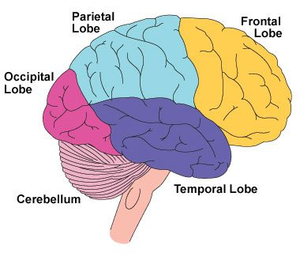
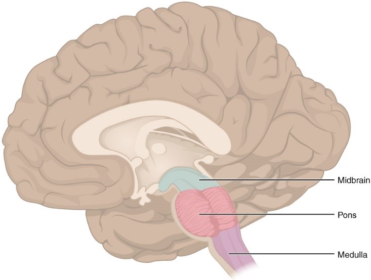
The cerebellum is in the posterior aspect of the brain, under the tentorium cerebelli.
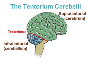
It is located in the posterior cranial fossa (the base of your skull, more specifically, the floor of your cranial cavity). It contains 50% or more of the neurons in your brain, so it’s a very important structure in your brain. We’ll touch more upon the structure of the cerebellum, and then how that connects to its function.
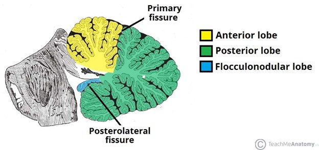
Structurally, the cerebellum has the anterior and posterior lobe, and a flocculonodular lobe.
There are folia and arbor vitae in the cerebellum. The folia make up the grey matter on the cerebellar cortex (surface), and the arbor vitae is the branched, white matter in the cerebellum. To explain grey and white matter, let me discuss neurons in detail.
A neuron is a cell that looks like this:
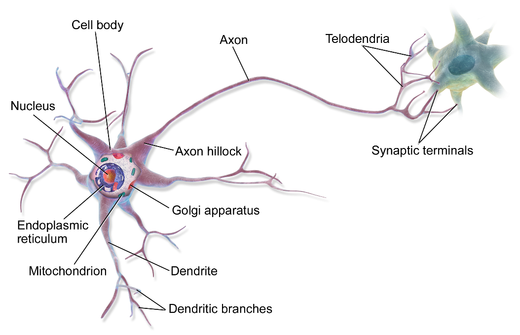
Lots of neuronal cell bodies make up the grey matter we see in the cerebellum. Lots of axons make up the white matter of the arbor vitae. Axons are covered in a fatty substance called myelin, and this myelin looks white. That’s why it’s called white matter.
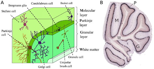
In the cerebellum, there are three important layers to remember. These are the granular, purkinje, and molecular cell layer. The molecular cell layer is at the very surface of the cerebellum, then comes the purkinje layer, and finally the granular layer is further inside the cerebellum.
Now, I could jump straight into the neuroscience and bombard you with complicated words that make no sense, but how would that help you?
Let’s try to simplify it, and just discuss movement first.
Let’s say that I want to move. I go to my friend called the primary motor cortex. I tell the primary motor cortex to start up a movement. The primary motor cortex will send a signal from my brain down to my spinal cord. A neuron projecting from my spinal cord will stick to a muscle in my arm, torso, or leg, and get me to move.
Here’s a diagram:
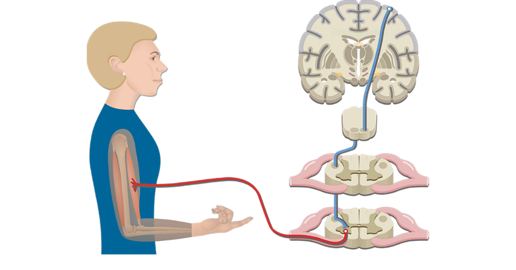
So, my brain sends a signal down my spinal cord to my arm (bicep), and I can move. Try to get that to sink in first.
Let’s try to get more specific.
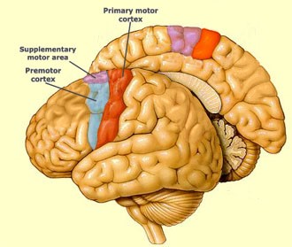
Now, we’re going to introduce what plans out you motor movements, the supplementary motor area and the premotor cortex. The supplementary motor area and premotor cortex have neurons that branch to the primary motor cortex. The supplementary motor area and premotor cortex are responsible for planning your movements and sending signals to your primary motor cortex.
The primary motor cortex is filled with neurons that allow sodium and potassium ions along with other nutrients through their cell membranes. The movement of this fluid allows signals to be sent throughout your body, via your neurons. Neurons in your motor cortex continue sending signals down the brain through their axons and then these axons project to your medulla oblangata.
At your medulla oblangata these axons cross over to the other half of your body, and then project to another neuron in your spinal cord.
Due to this, the left side of your brain controls the movement of the right side of your body, and the right side of your brain controls the movement of the left side of your body.
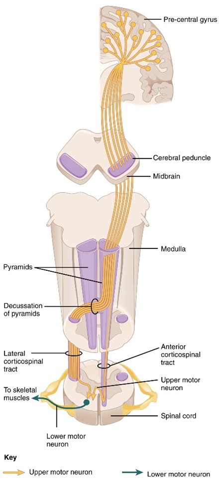
We call the neuron in your primary motor cortex an upper motor neuron because it is in your brain, and the neuron connecting to your muscle a lower motor neuron. The lower motor neuron connects to a muscle via a neuromuscular junction, sending the chemical acetylcholine to muscle cells, causing them to be able to contract.
For brevity’s sake I won’t go deeper into what occurs in a muscular contraction, synapse, and action potentials, but I’ll leave some resources at the end of the article you can use to learn more about that.
Now you know that the primary motor cortex is responsible for generating movements in your body. However, I still haven’t told you about the role of the cerebellum.
The cerebellum will usually be sending chemicals to your primary motor cortex telling it not to move.
Your cerebellum has three important nuclei, which are just collections of neuronal cell bodies. These are called the fastigial, interposed, and dentate nucleus. We will only discuss the dentate nucleus.
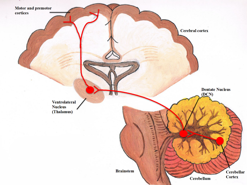
The dentate nucleus will send a chemical called gamma-Aminobutyric acid (GABA) to the ventrolateral nucleus of the thalamus. GABA is an inhibitory chemical. If you look at the diagram above, trace your finger from the red line extending the dentate nucleus to the ventrolateral nucleus of the thalamus. This is the important pathway to remember when we discuss the thalamus. It is called the dentatothalamocortical pathway.
In the dentatothalamocortical pathway, an axon projects from the dentate nucleus to the ventrolateral nucleus of the thalamus. GABA (inhibitory chemical) is released by the axon, causing the thalamus not to sends a chemical called glutamate (excitatory chemical) to the primary motor and premotor cortices.
So, if the thalamus cannot send the excitatory glutamate to the primary motor and premotor cortices, you cannot move. Your primary motor cortex isn’t receiving that exciting signal that tells it that it needs to allow a movement to happen.
However, that still doesn’t tell you anything about how the cerebellum allows you to make smooth movements. Don’t worry, we’ll get to that.
If you want to be able to move, it is clear that we have to make sure your dentate nucleus is not sending that inhibitory GABA to your thalamus, so your thalamus can be activated. However, how does that happen? Let’s go over the details.
You have a bunch of neurons that take care of the sensory information that comes into your body. The prickling you feel on your arm, the sounds you hear, the sights you see, all of those sensations are processed by neurons. Even the sensory information about where you are in space. Your muscles and joints are always sending information up to your cerebellum, so that your cerebellum knows where you are.
A sensory neuron will send this information (about where you are in space, remember proprioception) up to your cerebellum via climbing or mossy fibers. Climbing fibers are given that name due to the way they “climb up” purkinje cells. Mossy fibers were named mossy because—well they look like they’re covered in moss.
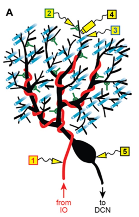
Your climbing fibers are the orange, and the mossy fibers are in blue. The purkinje cell is the thing that looks like a black bean with black lines extending out of it https://doi.org/10.3389/fncir.2013.00115
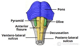
Climbing fibers come from a part of your brain called the olive.
Mossy fibers come from a myriad of places, like your face, limbs, and red nucleus. Just remember that they carry information about your body position.
Climbing and mossy fibers send excitatory signals to the purkinje and granule cells, respectively.
Granule cells then send excitatory signals to the purkinje cells, via the parallel fibers.
Purkinje cells then send inhibitory signals (GABA) to the deep cerebellar nuclei (fastigial, interposed, dentate). This is how the dentate nucleus is inhibited.
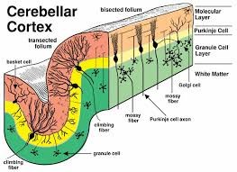
Try to figure out what is going on in this diagram :)
Your cerebellum will be constantly sending signals to your primary motor cortex, judging whether and how much your muscles should move. It is quite a fascinating component of the brain.
The cerebellum takes information about joint position, muscle tone, and muscle tension. It then allows you to make smooth, coordinated movements.
Hope you enjoyed the article and learned a little bit more about your cerebellum.
- Corticospinal Tract: https://youtu.be/9BaWBGRVxp8
- Action Potential: https://youtu.be/iBDXOt_uHTQ
- Neuromuscular Junction: https://youtu.be/zbo0i1r1pXA
- Muscular Contraction: https://youtu.be/BVcgO4p88AA
Sources:
- Jimsheleishvili S, Dididze M. Neuroanatomy, Cerebellum. [Updated 2021 Jul 31]. In: StatPearls [Internet]. Treasure Island (FL): StatPearls Publishing; 2021 Jan-. Available from: Link
- Richard S. Snell - Clinical Neuroanatomy